Conference Schedule
Day1: August 13, 2018
Keynote Forum

09:40-10:25
Konstantin Maksin
University of Information Technology and Management, Poland
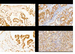
Title: Papillary thyroid cancer pathology-The modern challenge for multiprofile research teams
09:40-10:25
Biography
Abstract

10:25-11:10
10:25-11:10
Biography
Abstract
Tracks
- Microbial Biotechnology | Digital Pathology Exploring Scope & Application | Bacteriology & Virology | Artificial Intelligence & Computation | Microbiology research and advancement | Digital Image Analysis | Veterinary Microbiology
Location: Goya
Sinisa Vidovic
University of Minnesota, USA
Chair
11:30-12:05
Biography
Abstract
12:05-12:40
12:05-12:40
Biography
Abstract
13:55-14:30
Soumya Palliyil
University of Aberdeen, UK
Title: Protective effect of quorum quenching monoclonal antibodies in lethal Pseudomonas infection
13:55-14:30
Biography
Abstract
14:30-15:05
Kamlesh R Patel
Gujarat University, India
Title: How Digital pathaology can improve Healthcare in Semiurban and Rural areas
14:30-15:05
Biography
Kamlesh R Patel has completed his MD (Path and Bact) from Ahmedabad(GUJARAT University, India) in 1983. He was the Head of the department ofHaematology for two years, at GCRI. He has also worked in HisptopathologyDepartment at GCRI. He is serving as an chief Pathologist at Nakoda DiagnosticsAnd Research Center from last eight years and Managing 6 remotebranches with his two of assistant pathologist.
Abstract
15:05-15:40
Swee Hua Erin Lim
Abu Dhabi Womens College, UAE
Title: Ecotourism: Risk factor for transmission of MDR bacteria from non-human primates?
15:05-15:40
Biography
Abstract
15:40-16:15
Krzysztof Olesiejuk
Medical University of Lodz, Poland
Title: Impact of transcription factors on dendritic cell development
15:40-16:15
Biography
Abstract
Day2: August 14, 2018
Keynote Forum
10:00-10:45
Anshoo Agarwal
Northern Border University, Saudi Arabia
Title: Artificial intelligence and computational pathology: Are they future enemies of a pathologist?
10:00-10:45
Biography
Abstract
10:45-11:30
Ichiro Mori
International University of Health and Welfare, Japan
Title: Building international WSI telepathology full double-check system between Japan and Vietnam
10:45-11:30
Biography
Abstract
Tracks
- Microbial Biotechnology | Digital Pathology Exploring Scope & Application | Bacteriology & Virology | Artificial Intelligence & Computation | Microbiology research and advancement | Digital Image Analysis | Veterinary Microbiology
Location: Goya

Anshoo Agarwal
Northern Border University, Saudi Arabia
Chair

12:50-13:25
Anshoo Agarwal
Northern Border University, Saudi Arabia
Title: Role of robotic telepathology for frozen-section diagnosis
12:50-13:25
Biography
Abstract
14:25-15:00
Helmi Mardassi
Pasteur Institute of Tunis, Tunisia
Title: An automated IS6110-based fingerprinting method for accurate estimation of recent tuberculosis transmission
14:25-15:00
Biography
Abstract